Table of Contents
- Introduction
- Definition
- Etiology
- Anatomy Recall
- Figure 1: Vestibular nuclei and Vestibular tracts
- Table 1: Functions and connections of Vestibular nuclei
- Pathophysiology
- Clinical Evaluation
- History
- Duration of symptoms
- Trigger factors
- Table 2: Trigger factors of several causes of vertigo
- Associated symptoms
- Prior medical history
- Physical Examination
- Head impulse, nystagmus, and test of skew (HINTS) plus test
- Table 3: HINTS plus test summary
- Figure 2: Head Impulse Test
- Dix-Hallpike maneuver
- Hearing test
- Additional neurologic signs
- Diagnostic Tests
- Neuroimaging
- Additional studies
- Approach
- Figure 3: Approach to Vertigo in the Emergency Room
- Further Reading
- Bibliography
Primary Category
Essential Neurology
P-Category
Essential Neurology
Secondary Category
S-Category
Authors:


Introduction
- Vertigo is a false feeling of motion caused by dysfunction of the inner ear or the central vestibular system.
- This is a common complaint from patients in a primary care setting and emergency room.
- A thorough neurological examination and history taking are crucial to narrow down the diagnosis.
- Physicians need to apply the correct bedside testing to the right patient to avoid unnecessary neuroimaging.
Definition
- Vertigo is a symptom of vestibular dysfunction, typically described as an illusion of motion.
- Patients may find it hard to put their feelings into words. The most common complaint is a spinning sensation. Terms such as "whirling,” "tilting,” or "moving” are usually used.
Dizziness is an umbrella term for vertigo, pre-syncope, and disequilibrium. Physicians should confirm the symptom of vertigo before evaluating further.
Etiology
- Central lesion: caused by central vestibular system (e.g., cerebellum, brainstem, vestibular nuclei) lesions or dysfunction
- Cerebrovascular diseases (e.g., transient ischemic attack, ischemic and hemorrhagic stroke)
- Cerebellopontine angle tumor
- Migraine
- Multiple sclerosis
- Peripheral lesion: caused by the inner ear or the vestibulocochlear nerve dysfunction
- Benign positional paroxysmal vertigo (most common)
- Vestibular neuronitis
- Acute labyrinthitis
- Cholesteatoma
- Herpes zoster oticus (Ramsay Hunt syndrome)
- Ménière’s disease
- Otosclerosis
- Perilymphatic fistula
- Miscellaneous
- Cervical vertigo
- Drug-induced vertigo
- Psychological
Anatomy Recall
Figure 1: Vestibular nuclei and Vestibular tracts
.png)
- Primary sensory neurons in the vestibular ganglia send information about angular and linear motion from the semicircular canals and otolith organs (utricle and saccule), respectively, via the vestibular component of CN VIII to the vestibular nuclei.
- Vestibular nucleus complex:
- Four nuclei on each side of the brainstem.
- It lies on the lateral floor of the fourth ventricle in the pons and rostral medulla.
Table 1: Functions and connections of Vestibular nuclei
Vestibular Nuclei | Gives rise to | Connects with |
Lateral (Deiter’s) | Lateral vestibulospinal tract | ㅤ |
Superior | ㅤ | Medial longitudinal fasciculus |
Medial | Medial vestibulospinal tract | Medial longitudinal fasciculus |
Inferior | Medial vestibulospinal tract | ㅤ |
- The lateral and medial vestibulospinal tract descends via the spinal cord to innervate antigravity and head/neck muscle, respectively, maintaining balance and head position.
- The connection with the medial longitudinal fasciculus (MLF) creates the crosstalk between the vestibular and the visual system, displayed by the vestibulo-ocular reflex (VOR).
There are commissural fibers originating from vestibular nuclei passing through the vomiting center, which may explain why patients usually experience nausea during an acute vertiginous attack.
Pathophysiology
- An uncomfortable sensation (e.g., disequilibrium, vertigo) happens when the cerebellum detects a mismatch between the external feedback (e.g., spatial orientation information transmitted by the vestibular system) and its internal parameter.
- A lesion occurs anywhere along with the vestibular pathway → the cerebellum receives uneven information from both sides → the mismatch occurs → vertigo.
Clinical Evaluation
When approaching a patient with vertigo, remember the mnemonic “TITRATE”: TIming, TRiggers, And Targeted Examination.
History
Duration of symptoms
- Vertigo never continues permanently
- Many factors help the vestibular system compensate for the lesion over weeks and months.
- Constant “vertigo” lasting for months or years might not be vestibular.
- Due to the difficulty in describing vertiginous sensations, the clinical course is better than the quality of symptoms as a starting point for downstream diagnostic reasoning.
Caution: Some patients may claim they experience a consistent feeling of dizziness, but in fact, they are vulnerable to frequent episodic vertigo.
Trigger factors
- Head movement always exacerbates vertigo, and that is why patients avoid moving during a vertiginous attack.
If head movement does not worsen the attack, then such an episode might be another type of dizziness.
Table 2: Trigger factors of several causes of vertigo
Conditions | Trigger factors | Mechanism |
Benign Paroxysmal Positional Vertigo (BPPV) | Positional changes, particularly getting in or out of bed or turning over in bed | Dislodge calcium carbonate structures of the otolith organs into semicircular canals → disrupts the endolymph dynamic → spinning sensation |
Vestibular neuritis | Viral infection, especially upper airway infection | Postviral inflammation of the eighth cranial nerve |
Vertebral artery dissection | Neck hyperextension | ↓ blood supply to the posterior circulation → resembles lateral medullary dysfunction |
Perilymphatic fistula or Superior canal dehiscence | Coughing, exertion, or loud noises (Tullio phenomenon) | ↑ pressure transmitted from the cerebrospinal fluid (CSF) space to the inner ear via an abnormal anatomical defect |
Migrainous vertigo | Any trigger factors of migraine (e.g., flashing lights, sleep disturbance, certain food) | Some authors theorized that the neuropeptide surge causes vertigo, not the headache itself |
Associated symptoms
Nausea/vomiting
- Very common with an acute vertigo attack.
- It may cause dehydration and electrolyte imbalance in severe cases.
- Can present with both central and peripheral lesions.
Postural and gait instability
- The vestibular nuclei send signals to antigravity muscles via the vestibulospinal tract → uneven descending stimulations cause posture instability.
- Central lesions have a greater effect on balance probably because other pathways controlling posture are involved as well.
Drop attacks
- A sensation of being pulled to the ground while walking or standing without losing consciousness.
- Can be of cardiac, psychological, or vestibular origin.
- If patients present with vestibular dysfunction and a drop attack, they are very likely to have Ménière's disease.
Prior medical history
- Necessary to narrow down possible causes and save time in the diagnostic process.
- Risk factors such as hypertension, diabetes mellitus, smoking, and a history of vascular disease increase the suspicion of stroke.
- Past head trauma may cause perilymphatic fistula or BPPV.
- Prior cancer treatment with certain medications (e.g., cisplatin, aminoglycosides) suggests vestibular toxicity.
Physical Examination
Head impulse, nystagmus, and test of skew (HINTS) plus test
- Indication: Patients with continuous vertigo (lasting for hours/days), ongoing vertigo, and spontaneous nystagmus.
Misuse of the HINTS exam may lead to false-negative (e.g., stroke) or false-positive results (e.g., BPPV, vestibular migraine).
Table 3: HINTS plus test summary
VOR: Vestibulo-ocular reflex; HIT: Head Impulse Test; AICA: Anterior inferior cerebellar artery
ㅤ | Goal | Maneuver | Results | Interpretation | Note |
Head Impulse Test | Evaluate the VOR | 1. Ask a patient to fixate on a stationary object in front of them (e.g., a physician’s nose, a mask) | Normal: The patient maintains the eyes fixation during head rotation | Normal HIT suggests a central lesion (most likely stroke) | Remember to ask for a neck injury before performing the maneuver |
ㅤ | ㅤ | 2. Move the head off-center ~20-30 degrees | Abnormal: The patient cannot maintain the eyes fixation during head rotation, followed by a corrective shift of the eyes back to the stationary target (correction saccade) | Abnormal HIT mostly suggests a peripheral lesion (most likely vestibular neuritis) | Rotate the head from off-center toward the center, instead of doing the opposite, to avoid neck injury |
ㅤ | ㅤ | 3. Rapidly rotate the patient's head toward the center and assess their ability to maintain eyes fixation | ㅤ | ㅤ | ㅤ |
ㅤ | ㅤ | 4. For more details, watch the video: t.ly/dV-I | ㅤ | ㅤ | ㅤ |
Nystagmus | Evaluate spontaneous (primary) and gaze-evoked nystagmus | Look for the direction (left vs. right) of spontaneous horizontal nystagmus
| Unidirectional nystagmus: The direction of nystagmus does not change with gaze change
| The fast phase beats away from the lesion
| If the patient doesn’t have spontaneous nystagmus, then skip the HINTS plus test |
ㅤ | ㅤ | Look for gaze-evoked nystagmus while examining the extraocular muscles | Bidirectional nystagmus: The direction of nystagmus changes with gaze change | Bidirectional nystagmus indicates a central lesion | If the nystagmus is vertical, then skip the HINTS plus test and rapidly investigate for a central lesion |
ㅤ | ㅤ | For more details, watch the video: t.ly/zu6J | ㅤ | Unidirectional nystagmus indicates a peripheral lesion | ㅤ |
ㅤ | ㅤ | ㅤ | ㅤ | Alexander’s law: Nystagmus intensity ↑ when the patient looks toward the fast phase and ↓ when looking toward the slow phase | ㅤ |
Test of Skew | Evaluate the vertical ocular alignment | Alternate cover test:
1. The patient maintains a fixed central gaze and opened eyes during the examination
| Absent: The eyes remain in a fixed central gaze when uncovered | Vertical ocular misalignment (Skew deviation) suggests damage at the otolithic vestibular nuclei → central lesion, most likely located in the posterior fossa
| Do not touch the patient’s face during the maneuver, which triggers eye blinking → error evaluation |
ㅤ | ㅤ | 2. Repeatedly cover one eye, and then the other eye while looking for a vertical deviation from the central gaze upon uncovering the eye | Presented: The eyes A refixation saccade occurs upon uncovering the eyes → vertical ocular misalignment | ㅤ | ㅤ |
ㅤ | ㅤ | For more details, watch the video: t.ly/YGQ | Note: horizontal movement is not skew | ㅤ | ㅤ |
Finger Rub Test | The “plus” component of the traditional HINTS test
| 1. The examiner rubs their fingers together and asks if there is a decrease in hearing
| Normal: No hearing change from the patient’s baseline
| AICA supplies blood to the labyrinth of the inner ear, unlike other cerebellar arteries infarction, complete AICA infarction compromises VOR and causes hearing loss, resulting in abnormal HIT, and abnormal finger rub test [15] | ㅤ |
ㅤ | To screen for Anterior inferior cerebellar artery (AICA) infarct in patients who have abnormal head impulse test | 2. Compare both sides | Abnormal: a new and unequal hearing ability between two sides | ㅤ | ㅤ |
ㅤ | ㅤ | For more details, watch the video: t.ly/9Sdz | ㅤ | ㅤ | ㅤ |
Figure 2: Head Impulse Test

In lateral medullary stroke (or Wallenberg Syndrome), the ischemic lesion spares the superior and most part of the medial vestibular nuclei (main sources of the MLF) → intact vestibulo-ocular reflex (VOR) → normal head impulse test.
One study in a stroke center showed that the HINTS plus test had a higher sensitivity in detecting stroke than the ABCD² score.
Dix-Hallpike maneuver
- To confirm the diagnosis of Benign Paroxysmal Positional Vertigo (BPPV).
Although Dix-Hallpike is a simple and reliable bedside test, emergency physicians often overlook or misuse it.
- We will discuss Dix-Hallpike and similar maneuvers in more detail in our BPPV chapter.
- To learn how to perform the test, we suggest this video:

Hearing test
- To further investigate the cause of vertigo.
- The examiner can check the hearing ability quickly with several methods (eg, whisper to the ear, finger rub).
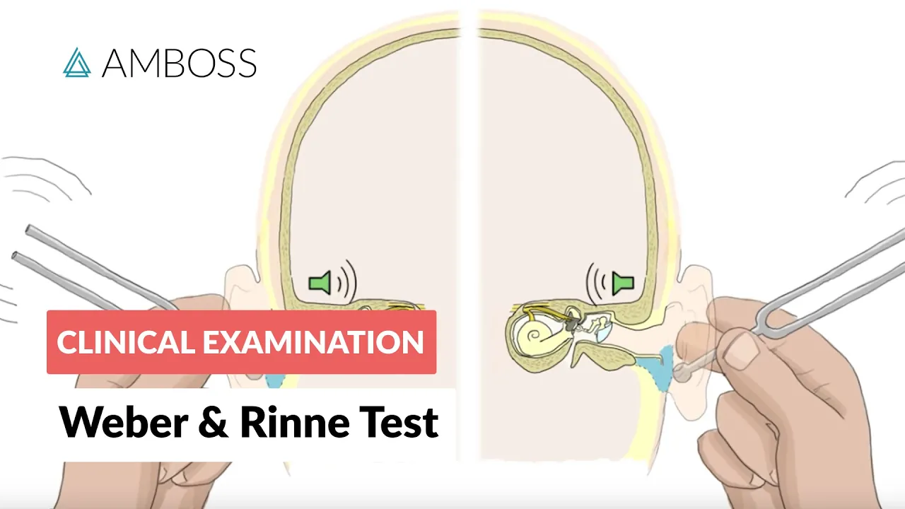
- The Weber and Rinne tests are used to distinguish conductive and sensorineural hearing loss. We suggest this video to learn how to perform and interpret Weber and Rinne test.
Additional neurologic signs
A careful neurologic examination is a must in any patient presenting with vertigo. Physicians should assess all cranial nerves, muscle strength, sensory function, coordination, gait, and deep tendon reflexes.
- Focal neck pain after trauma or spontaneously appearing may suggest vertebral artery dissection.
- Lateral medullary signs (e.g., diplopia, dysarthria, dysphagia, focal weakness, or paresthesias) suggests brainstem infarction.
- Signs of local demyelination in patients with multiple sclerosis.
- Some patients experience a sensation of aural fullness during an attack of Meniere’s disease.
- Patients may experience headache, photophobia, and sonophobia in vestibular migraine, but not in all attacks.
The Five Dangerous D's: Dysphagia, Dysarthria, Diplopia, Dysmetria, and Dysphonia strongly suggest a central cause of vertigo.
Diagnostic Tests
Neuroimaging
- Indicated when suspecting vertigo secondary to a central lesion.
- The procedure of choice in most cases is magnetic resonance imaging (MRI) of the brain with or without a magnetic resonance angiogram (MRA).
MRI yields a better diagnostic value than computed tomography (CT) scan in evaluating the posterior fossa lesion and acute ischemic changes.
MRA shows specificity and sensitivity of over 95 percent in detecting stenosis or occlusion of the posterior circulation.
Additional studies
- Laboratory tests: considered based on history and clinical presentation
Laboratory tests only detect the cause of vertigo in <1% of patients with all kinds of dizziness.
- Electrocardiogram (ECG): to look for evidence of cardiogenic syncope or arrhythmia.
- Audiometry
- More sensitive than office testing (e.g., Weber and Rinne test, finger rub test) to identify hearing loss.
- Quantify the hearing loss at high and low frequencies → to identify low-frequency sensorineural hearing loss → necessary to confirm Meniere’s disease.
- Can detect unilateral hearing loss in almost all cases of vestibular schwannoma and may be a powerful screening test for this tumor.
- Caloric testing: to assess isolated peripheral vestibulopathy
- Less accurate than the head impulse test in acute settings.
- However, the caloric test does not require head movement → favorable in patients with limited cervical mobility or coma.
- To further understand and learn how to perform the test, watch the video: t.ly/_WQa

Approach
- The differential diagnosis from a complaint of vertigo is massive; therefore, a diagnostic workup for a patient should be individualized.
- However, in patients with an acute onset of vertigo, we should initially rule out stroke, especially in the emergency room environment.
Figure 3: Approach to Vertigo in the Emergency Room

- To understand the above approach in more detail, we suggest watching this video from Dr. Johns - the author of the flow chart:

Further Reading
- Stanton, M., & Freeman, A. M. (2022). Vertigo. In StatPearls. StatPearls Publishing.
- Muncie, H. L., Sirmans, S. M., & James, E. (2017). Dizziness: Approach to Evaluation and Management. American family physician, 95(3), 154–162.
- Dr. Johns’s Youtube Channel: https://www.youtube.com/c/PeterJohns
Bibliography
- Zajonc, T. P., & Roland, P. S. (2005). Vertigo and motion sickness. Part I: vestibular anatomy and physiology. Ear, nose, & throat journal, 84(9), 581–584.
- Newman-Toker, D. E., & Edlow, J. A. (2015). TiTrATE: A Novel, Evidence-Based Approach to Diagnosing Acute Dizziness and Vertigo. Neurologic clinics, 33(3), 577–viii. https://doi.org/10.1016/j.ncl.2015.04.011
- Lacour, M., Helmchen, C., & Vidal, P. P. (2016). Vestibular compensation: the neuro-otologist's best friend. Journal of neurology, 263 Suppl 1, S54–S64. https://doi.org/10.1007/s00415-015-7903-4
- Newman-Toker, D. E., Cannon, L. M., Stofferahn, M. E., Rothman, R. E., Hsieh, Y. H., & Zee, D. S. (2007). Imprecision in patient reports of dizziness symptom quality: a cross-sectional study conducted in an acute care setting. Mayo Clinic proceedings, 82(11), 1329–1340. https://doi.org/10.4065/82.11.1329
- Stanton, V. A., Hsieh, Y. H., Camargo, C. A., Jr, Edlow, J. A., Lovett, P. B., Goldstein, J. N., Abbuhl, S., Lin, M., Chanmugam, A., Rothman, R. E., & Newman-Toker, D. E. (2007). Overreliance on symptom quality in diagnosing dizziness: results of a multicenter survey of emergency physicians. Mayo Clinic proceedings, 82(11), 1319–1328. https://doi.org/10.4065/82.11.1319
- Kim J. S. (2020). When the Room Is Spinning: Experience of Vestibular Neuritis by a Neurotologist. Frontiers in neurology, 11, 157. https://doi.org/10.3389/fneur.2020.00157
- Cutrer, F. M., & Baloh, R. W. (1992). Migraine-associated dizziness. Headache, 32(6), 300–304. https://doi.org/10.1111/j.1526-4610.1992.hed3206300.x
- Kerber, K. A., Brown, D. L., Lisabeth, L. D., Smith, M. A., & Morgenstern, L. B. (2006). Stroke among patients with dizziness, vertigo, and imbalance in the emergency department: a population-based study. Stroke, 37(10), 2484–2487. https://doi.org/10.1161/01.STR.0000240329.48263.0d
- Baloh R. W. (1998). Differentiating between peripheral and central causes of vertigo. Otolaryngology--head and neck surgery : official journal of American Academy of Otolaryngology-Head and Neck Surgery, 119(1), 55–59. https://doi.org/10.1016/S0194-5998(98)70173-1
- Lee, H., Yi, H. A., Lee, S. R., Ahn, B. H., & Park, B. R. (2005). Drop attacks in elderly patients secondary to otologic causes with Meniere's syndrome or non-Meniere peripheral vestibulopathy. Journal of the neurological sciences, 232(1-2), 71–76. https://doi.org/10.1016/j.jns.2005.01.012
- Kutlubaev, M. A., Xu, Y., Manchaiah, V., Zou, J., & Pyykkö, I. (2022). Vestibular drop attacks in Ménière's disease: A systematic review and meta-analysis of frequency, correlates and consequences. Journal of vestibular research : equilibrium & orientation, 32(2), 171–182. https://doi.org/10.3233/VES-201514
- Fitzgerald D. C. (1995). Persistent dizziness following head trauma and perilymphatic fistula. Archives of physical medicine and rehabilitation, 76(11), 1017–1020. https://doi.org/10.1016/s0003-9993(95)81041-2
- Andersson, H., Jablonski, G. E., Nordahl, S., Nordfalk, K., Helseth, E., Martens, C., Røysland, K., & Goplen, F. K. (2022). The Risk of Benign Paroxysmal Positional Vertigo After Head Trauma. The Laryngoscope, 132(2), 443–448. https://doi.org/10.1002/lary.29851
- Gerlier, C., Hoarau, M., Fels, A., Vitaux, H., Mousset, C., Farhat, W., Firmin, M., Pouyet, V., Paoli, A., Chatellier, G., & Ganansia, O. (2021). Differentiating central from peripheral causes of acute vertigo in an emergency setting with the HINTS, STANDING, and ABCD2 tests: A diagnostic cohort study. Academic emergency medicine : official journal of the Society for Academic Emergency Medicine, 28(12), 1368–1378. https://doi.org/10.1111/acem.14337
- Lee, H., Kim, J. S., Chung, E. J., Yi, H. A., Chung, I. S., Lee, S. R., & Shin, J. Y. (2009). Infarction in the territory of anterior inferior cerebellar artery: spectrum of audiovestibular loss. Stroke, 40(12), 3745–3751. https://doi.org/10.1161/STROKEAHA.109.564682
- Lee, S. H., Kim, J. M., Schuknecht, B., & Tarnutzer, A. A. (2020). Vestibular and Ocular Motor Properties in Lateral Medullary Stroke Critically Depend on the Level of the Medullary Lesion. Frontiers in neurology, 11, 390. https://doi.org/10.3389/fneur.2020.00390
- Newman-Toker, D. E., Kerber, K. A., Hsieh, Y. H., Pula, J. H., Omron, R., Saber Tehrani, A. S., Mantokoudis, G., Hanley, D. F., Zee, D. S., & Kattah, J. C. (2013). HINTS outperforms ABCD2 to screen for stroke in acute continuous vertigo and dizziness. Academic emergency medicine : official journal of the Society for Academic Emergency Medicine, 20(10), 986–996. https://doi.org/10.1111/acem.12223
- Dmitriew, C., Bodunde, O., Regis, A., Lepage, R., Turgeon, Z., Johns, P., McIsaac, S., & Ohle, R. (2021). The use and misuse of the Dix-Hallpike test in the emergency department. CJEM, 23(5), 613–616. https://doi.org/10.1007/s43678-021-00110-1
- Knox, G. W., & McPherson, A. (1997). Menière's disease: differential diagnosis and treatment. American family physician, 55(4), 1185–1194.
- Lawhn-Heath, C., Buckle, C., Christoforidis, G., & Straus, C. (2013). Utility of head CT in the evaluation of vertigo/dizziness in the emergency department. Emergency radiology, 20(1), 45–49. https://doi.org/10.1007/s10140-012-1071-y
- Becker, K. J., Purcell, L. L., Hacke, W., & Hanley, D. F. (1996). Vertebrobasilar thrombosis: diagnosis, management, and the use of intra-arterial thrombolytics. Critical care medicine, 24(10), 1729–1742. https://doi.org/10.1097/00003246-199610000-00022
- Hoffman, R. M., Einstadter, D., & Kroenke, K. (1999). Evaluating dizziness. The American journal of medicine, 107(5), 468–478. https://doi.org/10.1016/s0002-9343(99)00260-0
- Selters, W. A., & Brackmann, D. E. (1977). Acoustic tumor detection with brain stem electric response audiometry. Archives of otolaryngology (Chicago, Ill. : 1960), 103(4), 181–187. https://doi.org/10.1001/archotol.1977.00780210037001
- Morrison, M., Korda, A., Zamaro, E., Wagner, F., Caversaccio, M. D., Sauter, T. C., Kalla, R., & Mantokoudis, G. (2022). Paradigm shift in acute dizziness: is caloric testing obsolete?. Journal of neurology, 269(2), 853–860. https://doi.org/10.1007/s00415-021-10667-7
AizaMD™: Revolutionizing Clinical Documentation
Discover the power of our ambient clinical documentation system, designed to transform clinical encounters into structured SOAP notes with unmatched ease. Experience exceptional value for less than $3 per day—cheaper than your daily coffee!
- Save Time: Free up over 90 minutes daily for each provider.
- Boost Revenue: Increase daily revenue by at least $1,000 per provider.
- Enhance Coding Quality: Our detailed documentation supports superior coding accuracy, ensuring optimal reimbursement.
- Maximize Engagement and Interaction: Dedicate more time to patient care and less to typing, fostering richer and more effective conversations between clinicians and patients


AizaMD™: Automated Radiology Report Generation!
Discover our breakthrough Radiology AI reporting platform built on Ambient AI. It enhances productivity and minimizes fatigue. Benefit from best-in-class accuracy with our automated radiology report generation, all at market-leading pricing.
📈 Efficiency: Cut dictation times by up to 50% (Less words, More report!
🎯 Focus: Keep your eyes on the images, not the keyboard!
💸 Revenue: Boost revenue by at least 20%
📑 Clarity: Patient summary in plain English


Written by